
Mammary tumors – Dogs

Mammary tumors are very common in female dogs, especially those that are not spayed or were spayed later in life. About half of mammary tumors are benign, while the other half are malignant (cancerous).
Fortunately, when a mass is detected early and removed before it spreads, surgery can be curative in many cases.
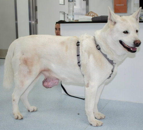
How to reduce the risk?
Hormones produced by the ovaries play an important role in the development of many mammary tumors. This is why early spaying is the best prevention method.
[ – ] Before the first heat: the risk of developing a mammary tumor is very low.
[ – ] After the first heat: the risk remains relatively low, but begins to increase.
[ – ] After several heat cycles: the protective effect of spaying becomes less important and the risk of mammary tumor increases.
The ideal age for spaying must also consider several other factors, including the breed, size, and health status of your dog. Your veterinarian can help you determine the best time for her.
How to detect an abnormality at home?
The risk of mammary tumor is higher in unspayed dogs or those spayed late. However, a mammary mass can also appear in a spayed dog.
Normal anatomy: A female dog usually has 10 mammary glands, arranged in two rows on the belly. They are not always perfectly symmetrical.
What to look for? By gently petting her belly, look for a new lump or a small firm mass under the skin. Mammary tumors appear more frequently in the glands located near the hind legs.
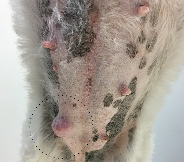
⚠ At what age to check? Mammary tumors are mostly diagnosed in middle-aged to older dogs. We recommend starting to regularly palpate the teats from the age of 6, whether she is spayed or not.
If you discover a mass, even a very small one, do not wait for it to grow before consulting. The smaller a tumor is when removed, the better the prognosis generally is.
A mass has been discovered: what is the next step?
Before removing a mammary mass, certain checks may be recommended to determine its nature and look for signs of spread.
The staging
When a mammary tumor is potentially cancerous, one of the places it can spread is the lungs. Chest X-rays are therefore generally recommended before surgery.
Three X-rays taken in different positions allow us to examine the lungs from several angles and increase our chances of detecting any metastases.
If signs of spread are discovered, it does not necessarily mean that no treatment is possible. However, the prognosis and goals of treatment change.Not every lump located near a teat is necessarily a mammary tumor. Some masses can originate from the skin or subcutaneous tissues.
A fine-needle aspiration allows a few cells to be collected using a small needle. This test can help us rule out certain other causes of masses, but it does not always allow us to determine with certainty whether a mammary tumor is benign or malignant.
Mass evaluation
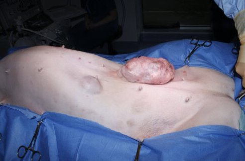 For this reason, when the mass can be completely removed, we often prefer an excisional biopsy: the mass is surgically removed, then sent to the laboratory for analysis. Tissue analysis, called histopathology, provides us with the definitive diagnosis and several important pieces of information about the prognosis.
[!] An exceptional case: Inflammatory Mammary Carcinoma (IMC)
This is a rare, but particularly aggressive form of breast cancer. Rather than forming a small, well-defined mass, it spreads into the skin’s tissues and lymphatic vessels. The teats can quickly become red, hot, thickened, swollen, and painful, which can sometimes look like an infection. This cancer spreads quickly and, unlike most other mammary tumors, surgery is generally not recommended. Treatment is primarily aimed at controlling pain and inflammation and preserving the animal’s quality of life.
What can the laboratory analysis reveal?
The analysis of the removed mass provides us with several important pieces of information. First, it determines what type of tumor it is and whether it is benign or malignant.
Benign tumors: They do not spread to other organs. When the mass is completely removed, no further treatment is usually necessary.
Malignant tumors: These are mammary cancers. Some progress slowly and can be cured by surgery, while others are more aggressive and have a higher risk of recurrence or spread.
When a tumor is cancerous, the laboratory also evaluates its grade, which tells us about its degree of aggressiveness. It looks for certain signs associated with a higher risk of spread, notably the presence of cancer cells in the blood or lymphatic vessels. Finally, it evaluates the surgical margins, meaning whether the tumor appears to have been completely removed.
What determines the prognosis?
Not all cancerous mammary tumors behave the same way. Several elements allow us to estimate their aggressiveness and the chances of a cure:
➤ Size: The smaller a mass is when removed, the better the prognosis generally is.
➤ Growth rate: A mass that grows quickly is generally more concerning.
➤ Appearance: Deeply attached, highly inflammatory, or ulcerated may be worrisome.
➤ Grade: A higher grade generally indicates a more aggressive tumor.
Spaying and Post-Surgery
In an unspayed dog, we may recommend spaying at the same time as the tumor removal. Spaying helps prevent future diseases (like pyometra) and could reduce the risk of new mammary tumors.
Once the mass is analyzed, the laboratory results will determine the next steps. For a benign tumor, no additional treatment is generally necessary.
For a malignant tumor, regular follow-up may be recommended (examination of teats, lymph nodes, X-rays). In some higher-risk cases, a consultation with an oncology specialist may be recommended.
To remember: it is best to have any new mass examined quickly.
We are by your side at every step. Do not hesitate to make an appointment!
